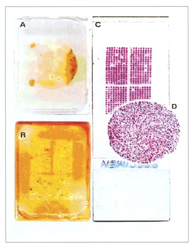
Figure 1.
Tissue microarray (TMA) manufacturing. A) Donor block from which several 0.6 mm tissue cores have been removed. Note that the original tissue block remains fully interpretable. B) Recipient block with the completed TMA. C) Hematoxylin & eosin stained tissue section of the TMA. D) Magnification of a tissue spot.

