Fig. 5.
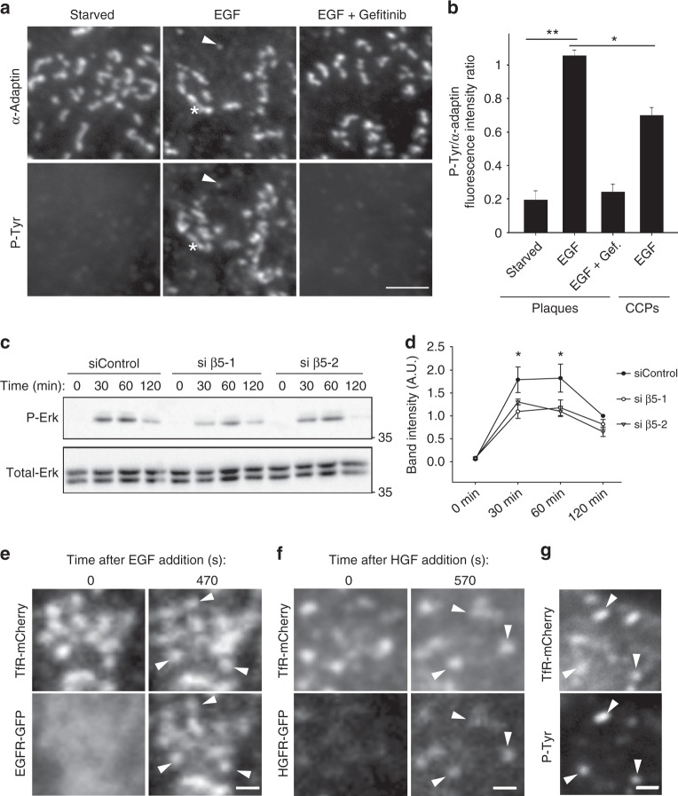
Clathrin-coated plaques locally regulate receptor-dependent Erk signaling. a HeLa cells seeded on collagen-coated glass were starved for 4 h and then treated or not with 10 ng/ml EGF for 5 min alone or added after 30 min preincubation with 10 µM Gefitinib. Cells were then fixed and stained for α-adaptin and phosphotyrosines. The arrowhead points to one CCP and the star marks a plaque. Scale bar: 2 μm. b Quantification of phosphotyrosines accumulation at plaques or CCPs in the indicated conditions. Results are expressed as mean ± SD (*P < 0.05, **P < 0.001, one-way analysis of variance—ANOVA. n = 3. Number of structures analyzed per condition: Starved 160 plaques; EGF: 452 plaques, EGF + Gefitinib: 301 plaques, CCPs/EGF: 200 pits). c Western-blot analysis of phospho-Erk (P-Erk) levels in starved HeLa cells treated with control or β5-specific siRNAs as indicated, and stimulated for the indicated time with 10 ng/ml EGF. Total-Erk was used as a loading control (representative image of three independent experiments). d Densitometry analysis of bands obtained in western-blots as in c. Results are expressed as mean ± SD (*P < 0.05, ANOVA. n = 3). e HeLa cells treated with β5-specific siRNAs were transfected with plasmids encoding for TfR-mCherry and EGFR-GFP and seeded on anti-mCherry antibodies-coated glass. Cells were serum-starved for 4 h and then treated with 10 ng/ml EGF for 470 s. Scale bar: 0.5 μm. Arrowheads point to plaques positive for EGFR-GFP. f HeLa cells treated with β5-specific siRNA were transfected with plasmids encoding for TfR-mCherry and HGFR-GFP and seeded on anti-mCherry antibodies-coated glass. Cells were serum-starved for 4 h and then treated with 50 ng/ml HGF for 570 s. Scale bar: 0.5 μm. Arrowheads point to plaques positive for HGFR-GFP. g HeLa cells treated with β5-specific siRNA were transfected with plasmids encoding for TfR-mCherry and seeded on anti-mCherry antibodies-coated glass. Cells were serum-starved for 4 h and then treated with 10 ng/ml EGF for 5 min prior to fixation and staining for phosphotyrosines. Scale bar: 1.5 μm. Arrowheads point to plaques positive for P-Tyr

