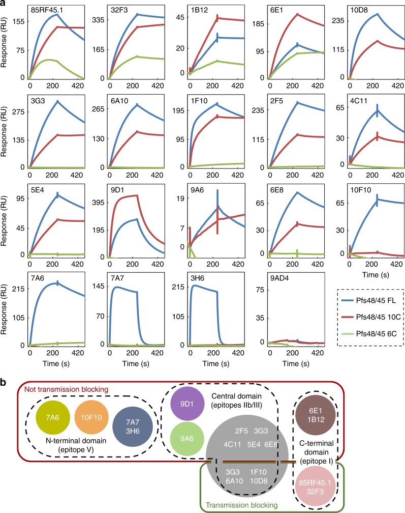
Fig. 3.
Mapping of anti-Pfs48/45 antibodies binding to different Pfs48/45 subdomains. a The indicated mAbs were immobilised on a Protein A/G chip at fixed concentration. Pfs48/45 FL (blue lines), Pfs48/45-10C (red lines) or Pfs48/45-6C (green lines) were then injected over the chip surface at fixed concentration. 9AD4 is a control antibody reactive against PfRH5. b Summary of epitope mapping experiments. Circles indicate the different competition groups among the mAbs, as shown in Fig. 2. mAbs are further grouped into transmission-blocking and non-transmission-blocking mAbs by green and red outlines, respectively. Black dashed outlines show which mAbs bind to the N-terminal, central (10C) or C-terminal (6C) domain. Previously defined epitopes I–V6, for anti-Pfs48/45 antibodies, are indicated

