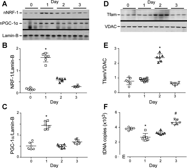
Fig. 4.
Lung mitochondrial biogenesis during S. aureus pneumonia. A: Western analyses of lung nuclear respiratory factor 1 (NRF-1) and peroxisome proliferator-activated receptor-γ coactivator-1α (PGC-1α) proteins showing nuclear accumulation of both proteins at day 1 postinfection. B and C: scatterplots of densitometry analysis of NRF-1 and PGC-1α. D: mitochondrial transcription factor A (Tfam) levels triple at day 2 postinoculation, and normalize by day 3. The Tfam reference is outer mitochondrial membrane protein VDAC. E: scatterplots of densitometry values for lung mitochondrial Tfam analysis. F: scatterplots for mitochondrial DNA (mtDNA) copy number in lung parenchyma after S. aureus infection. mtDNA copy number falls on day 1, recovers, and has increased by day 3. Bars show means ± SE for each group and time point; n = 6 mice per group; *P < 0.05 vs. day 0 control and #P < 0.05 vs. day 1 by one-way ANOVA.

