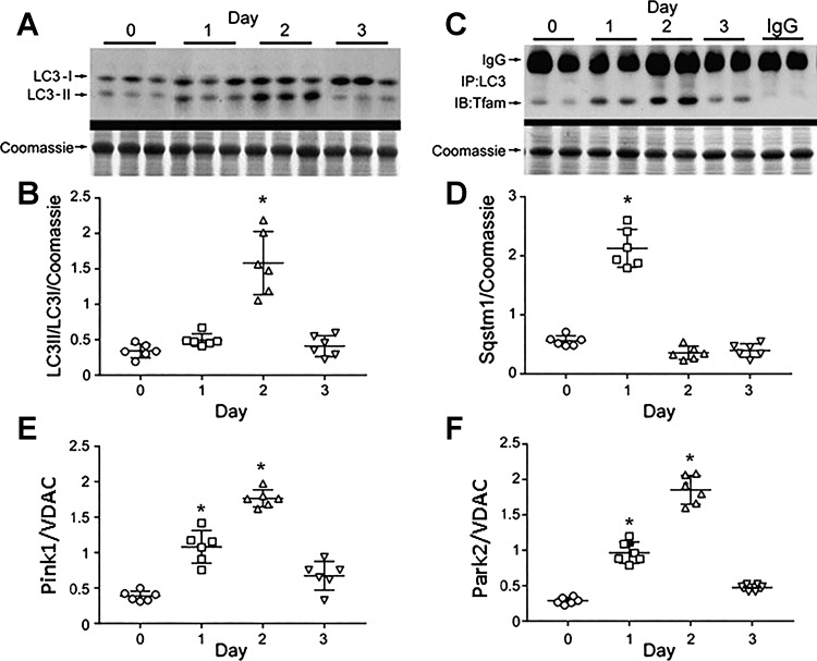
Fig. 5.
Mitophagy in S. aureus pneumonia. A: light chain 3B protein (LC3-I) cleavage to LC3II by Western blot pre-/post-S. aureus inoculation. B: scatterplots of lung autophagic index (LC3II/LC3I) pre-/post-S. aureus inoculation. The index increased at day 2. *P < 0.05 for n = 5 per group by two-way ANOVA. C: far Western immunoprecipitation of lung protein with anti-LC3 probed against Tfam. S. aureus inoculation enhances Tfam association with LC3. D: Western blot of lung Sqstm1 (p62) protein. S. aureus pneumonia enhances Sqstm1 levels at day 1 consistent with onset of mitophagy. E: scatterplots for Pink1 relative to VDAC pre-/postinoculation show higher Pink1 levels at days 1 and 2. F: scatterplots for Park2 mitophagy protein relative to VDAC pre-/postinoculation show higher Park2 (Parkin) at days 1 and 2. For scatterplots in D–F, bars are means ± SE for n = 6 per time point. *P < 0.05 vs. time 0 by one-way ANOVA.

