Figure 1.
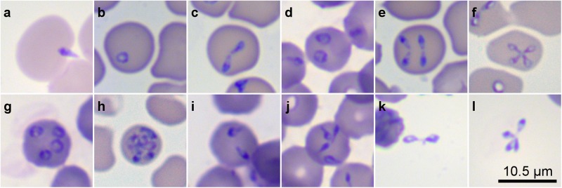
B. divergens forms inside and outside erythrocytes were identified in in vitro cultures by Giemsa staining. (a) A free merozoite invading a human erythrocyte. Panels b–j show different stages within the human erythrocyte. (b) Single round trophozoite. (c) Paired pyriforms, a stage formed by two attached pear-shaped sister cells. (d) Double round trophozoites, (e) Double paired pyriforms. (f) Tetrad (g). Quadruple round trophozoites. (h) Multiple parasites. (i) Double unattached pyriforms. (j) Quadruple unattached pyriforms. (k) Intact paired pyriform outside the erythrocyte. (l) Intact tetrad outside the erythrocyte. Slides were examined with a Primo Star microscope (Zeiss, Germany) at 100X magnification.

