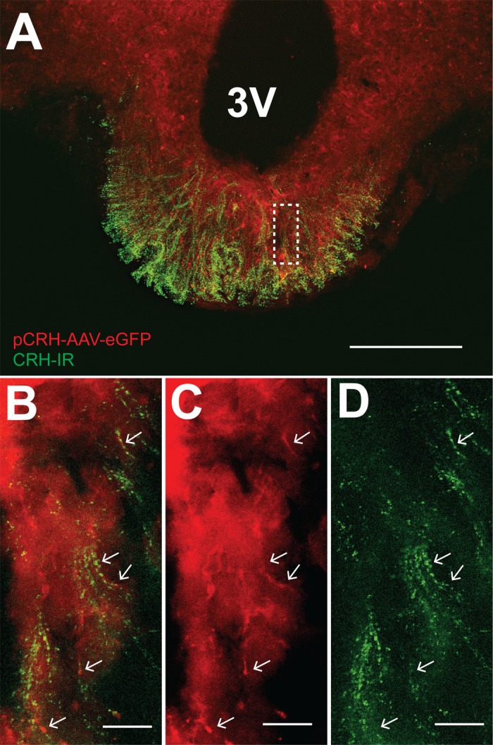
Fig. 8.

eGFP is expressed in CRH-IR fibers in the median eminence following transfection of the PVN with pCRH-AAV-eGFP. A: presumed CRH fibers from the PVN (pseudo-colored red) displayed CRH-IR (pseudo-colored green). Scale bar = 200 µm. B: higher-magnification image of region in dashed line box in A. Arrows indicate pCRH-AAV-eGFP fibers (C) coincident with CRH-IR (D). Scale bars for B–D = 15 µm.
