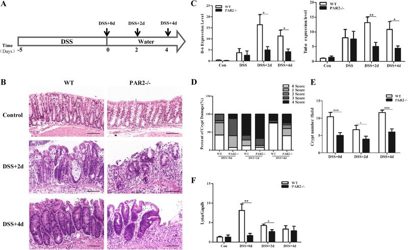
Fig. 1.
PAR2 deficiency impairs colonic regeneration following DSS-induced injury in mouse. a Schematic overview of DSS-induced colitis model in mouse. b H&E staining of colon sections taken from the mice 2 (DSS + 2d) or 4 days (DSS + 4d) after the administration of DSS. Scale bars represent 100 μm. c Expression of IL-6 and TNF-α mRNA were measured by real-time PCR with WT and PAR2-/-mouse colon. d Depth of crypt damage was assessed and shown (0, no damage; 1, basal 1/3; 2, basal 2/3; 3, only surface intact; and 4, entire crypt and surface loss). e Quantification of crypt number from five consecutive fields of each group. Data are shown as crypt number per field (magnification, × 250). f Expression of Ly6a mRNA was measured by real-time PCR with WT and PAR2-/- mouse colon. Data are shown as mean ± SEM. N.S., no significance; *p < 0.05; **p < 0.01; ***p < 0.001 vs. corresponding control

