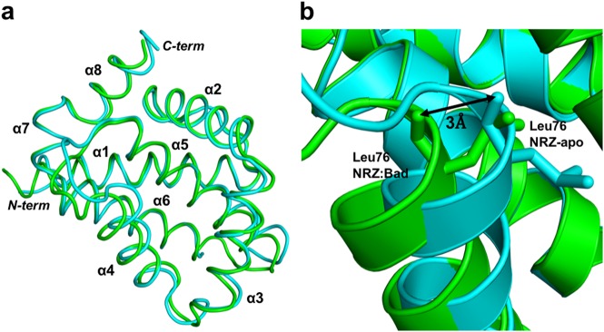
Fig. 3. Superimposition of apo NRZ with NRZ: zBad BH3 complex.
Comparison of the backbone structures of NRZ and NRZ: zBad BH3 complex. a Cartoon representation of apo NRZ (light blue) superimposed onto NRZ: zBad BH3 complex (green). The view is into the canonical hydrophobic binding groove formed by α2–5. b Close up view of NRZ helix α4, which is shifted by 3 Å from its original position in the apo NRZ structure upon zBad BH3 motif binding, thus enlarging the binding groove

