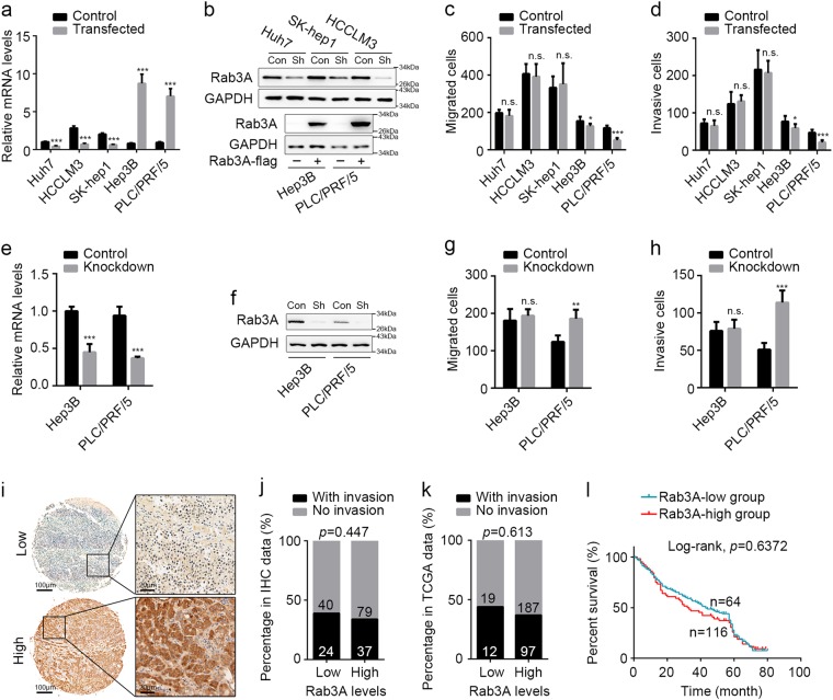
Fig. 2. Upregulated Rab3A malfunctions in most HCC cells.
a, b Efficiencies of Rab3A knockdown in Huh7 cells, SK-hep1 cells, and HCCLM3 cells, and Rab3A overexpression in Hep3B cells and PLC/PRF/5 cells were determined by qPCR and WB. GAPDH was used as internal control in both qPCR and WB analyses, and antibodies against Rab3A and GAPDH were used in WB. c, d Transwell assays for different HCC cells with Rab3A knockdown or overexpression. e, f Efficiencies of Rab3A knockdown in Hep3B cells and PLC/PRF/5 cells were determined by qPCR and WB. g, h Transwell assays for Hep3B and PLC/PRF/5 cells with Rab3A knockdown. i Representative IHC staining of tumor tissues with high or low levels of Rab3A in HCC. Regional magnification images were showed right. j, k Correlation between Rab3A expression and vessel invasion in IHC cohort and TCGA dataset. l Kaplan–Meier analysis for overall survival of HCC patients from IHC cohort. *p < 0.05, **p < 0.01, ***p < 0.001; NS, not significant

