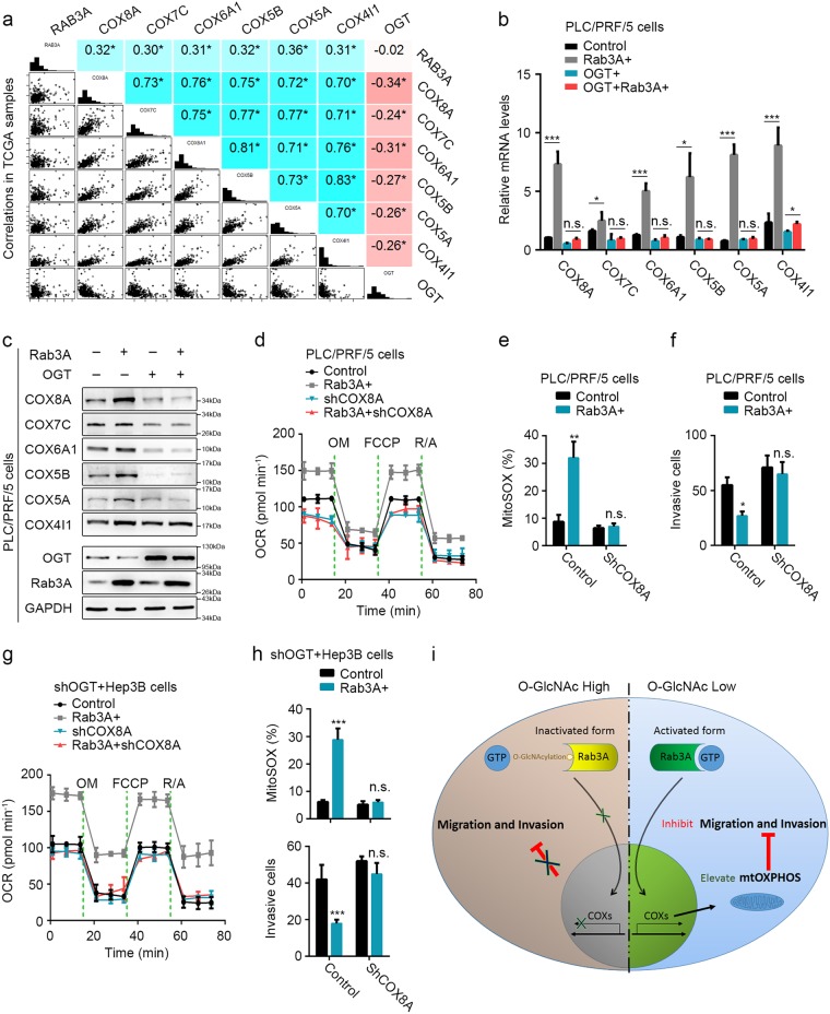
Fig. 7. COXs are critical for the functions of Rab3A and its O-GlcNAcylation in HCC.
a mRNA correlations of Rab3A, OGT, COX8A, COX7C, COX6A1, COX5B, COX8A, and COX4I1 in 442 primary HCC tumors of TCGA (n = 442 biologically independent patient samples). b, c mRNA and protein levels of COX8A, COX7C, COX6A1, COX5B, COX5A, and COX4I1 in PLC/PRF/5 cells with OGT and/or Rab3A expression. d The OCR curves in parental PLC/PRF/5, Rab3A + , shCOX8A, and Rab3A + shCOX8A cells treated with oligomycin, FCCP, and rotenone/antimycin A. e MitoSOX Red staining of PLC/PRF/5 cells with indicated treatments was analyzed by flow cytometry. f Transwell invasion assay for PLC/PRF/5 cells with indicated treatments. Six independent experiments were performed for each assay. g The OCR curves in OGT-knockdowned Hep3B, shOGT + Rab3A + , shOGT + shCOX8A, and shOGT + Rab3A + shCOX8A cells treated with oligomycin, FCCP, and rotenone/antimycin A. h MitoSOX Red staining of HeprB cells with indicated treatments was analyzed by flow cytometry, and the invasive ability was analyzed by transwell invasion assays under indicated treatments. i The schematic diagram describing the function of Rab3A and O-GlcNAcylation in HCC metastasis. Six independent experiments were performed for each assay. *p < 0.05, **p < 0.01, ***p < 0.001; NS, not significant

