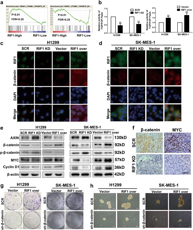
Fig. 5. RIF1 promotes cell growth and cancer stem cell-like phenotype in NSCLC by activating the Wnt/β-catenin pathway.
a GSEA plot showing that RIF1 expression was positively correlated with Wnt-activated gene signatures and inversely correlated with Wnt-suppressed gene signatures in the GEO data set (NCBI/GEO/GSE10245; n = 58). b The indicated cells were transfected with TOPflash or FOPflash and pRL-TK plasmids and then subjected to luciferase reporter assay 24 h post-transfection. The reporter activity was normalized to the activity of the pRL-TK reporter. c, d Subcellular β-catenin localization was detected in the indicated cells by immunofluorescence staining. Scale bar, 10 μm. e Western blot analyses of protein expression of AXIN, β-catenin, p-β-catenin, MYC, cyclin D1 and β-actin in RIF1-silenced or overexpressed H1299 and SK-MES-1 cells. f Representative images of IHC staining of the resected tumor. IHC analysis showed that RIF1 knockdown reduced the expression of β-catenin and MYC. g Colony formation assay of RIF1-overexpressed or control vector H1299 and SK-MES-1 cells with or without β-catenin knockdown. h Representative images of spheres formed by RIF1-overexpressed or control vector H1299 and SK-MES-1 cells with or without β-catenin knockdown. Scale bar, 100 μm. Data are expressed as means ± SD of three independent experiments. **P < 0.01

