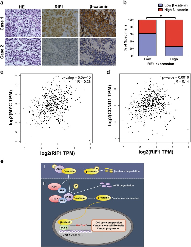
Fig. 8. Clinical relevance of RIF1 and Wnt/β-catenin signaling in NSCLC.
a, b Immunohistochemical staining suggesting that RIF1 expression correlated positively with β-catenin expression in 32 clinical NSCLC specimens. Percentage of NSCLC specimens showing low or high RIF1 expression relative to the expression levels of β-catenin. *P < 0.05. c, d Bioinformatics analysis based on GEPIA database showed the mRNA expression levels of RIF1 positively correlated with MYC (r = 0.28, P < 0.001) and CCND1 (r = 0.14, P = 0.0016) expression level in NSCLC tissues. e. Model of RIF1 promotes lung cancer progression and stem cell-like traits through PP1-mediated Wnt/β-catenin signaling. (I) In the absence of RIF1, AXIN–PP1 interaction is attenuated, leading to AXIN and β-catenin destruction complex stabilization, phosphorylation and degradation of β-catenin and thus attenuated Wnt signaling. (II) In the presence of RIF1, RIF1 guides PP1 to bind AXIN, resulting in AXIN dephosphorylation by PP1 and, ultimately its degradation. AXIN degradation leads to disassociation of the β-catenin destruction complex, an increase in β-catenin levels and in Wnt target gene transcription

