
FIG. 5.
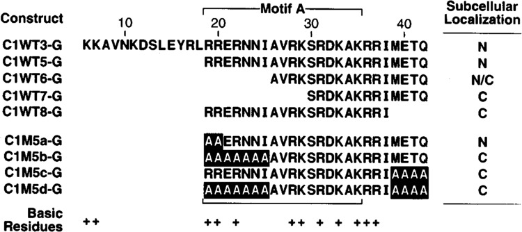
Mapping the N- and C-terminal boundaries of the CRP1 NLS. The subcellular location of β-gal fusion proteins was analyzed as described in Figs. 3 and 4. These mutants were constructed in the background of C1WT5G and are therefore designated by the number 5 followed by a lower case letter. M, mutated CRP1 sequence. N/C, protein appearing primarily in the nucleus but displaying light staining in the cytoplasm.