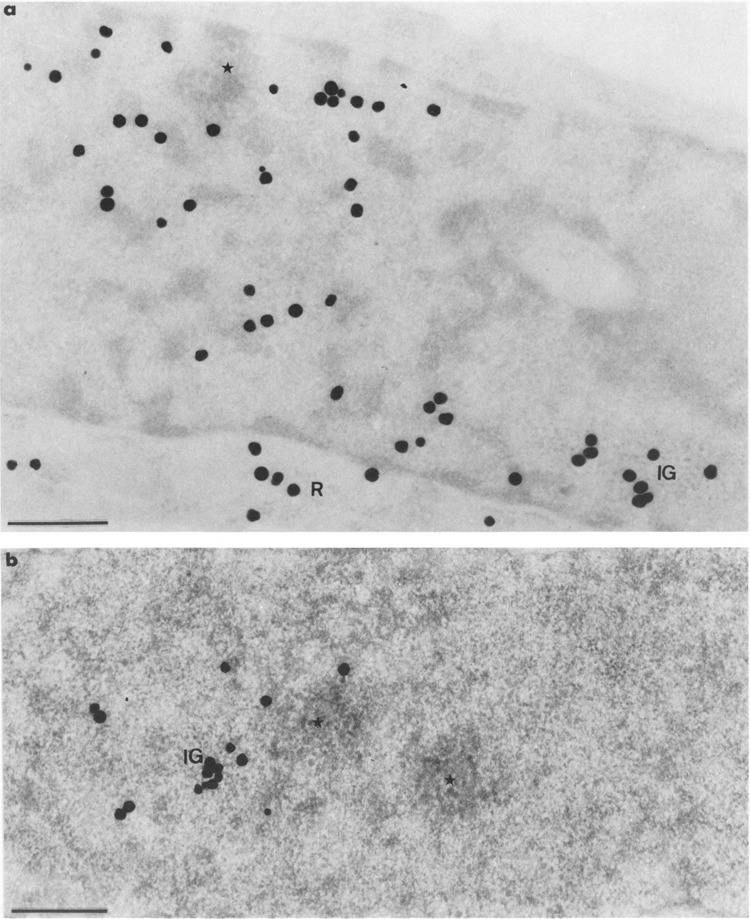
FIG. 10.

Autoradiograms of cells transiently expressing Us11 gene and pulse-labeled with tritiated uridine as in Fig. 8, then submitted to a chase of 60 min before fixation. Epon embedding. Bars represent 0.5 μm. (a) Uranyl acetate staining. The cluster of interchromatin granules (IG) is labeled whereas there is no labeling over the small focus of intermingled viral RNP fibrils (star). Silver grains are scattered over the remaining nucleoplasm and over the cytoplasmic ribosomes (R). (b) EDTA regressive staining. Part of nucleoplasm showing a labeled cluster of interchromatin granules (IG) and two unlabeled foci of intermingled RNP fibrils (stars), one of which being contiguous to the labeled cluster of interchromatin granules.
