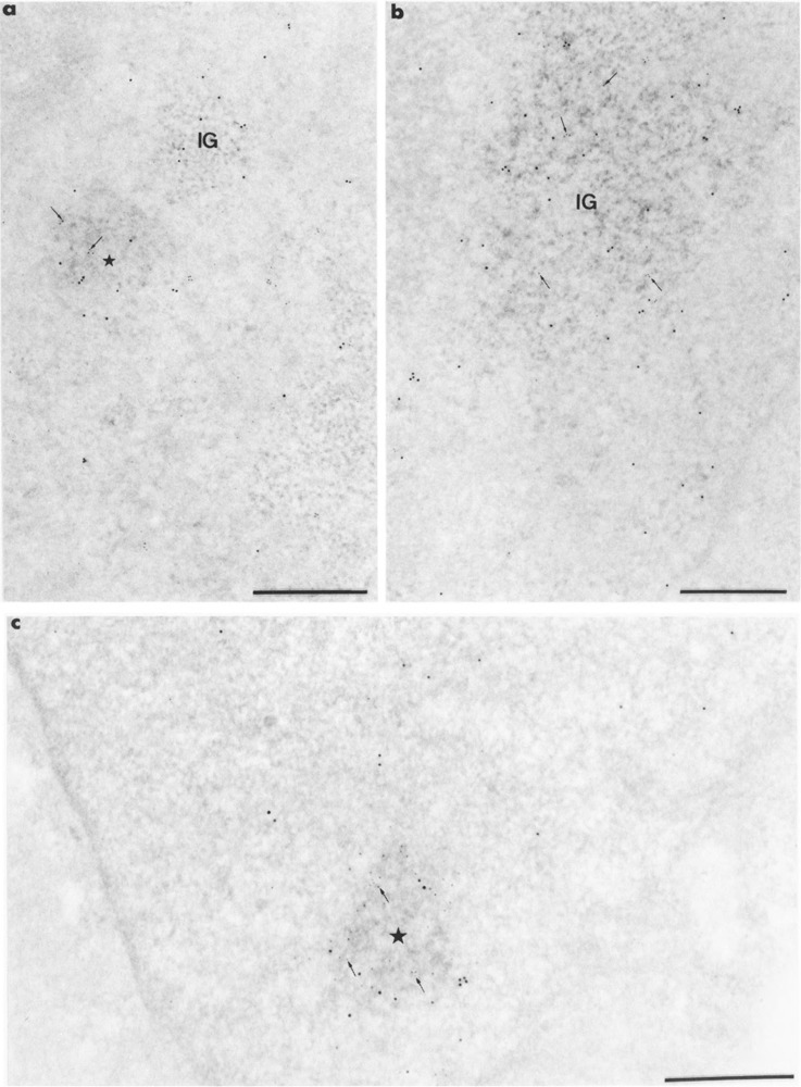
FIG. 7.

Colocalization of RNA polymerase II molecules and Us11 RNA on Lowicryl sections of cells transiently expressing Us11 gene. Formaldehyde in (a, b) and glutaraldehyde in (c) fixation. Uranyl acetate staining. Bars represent 0.5 μm. (a, b) mAb 7C2 and biotinylated Us11 DNA probe combination. Gold particles of 5-nm diameter label the protein whereas the 10-nm gold particles label the viral RNA. (a) Mixed 5-nm (arrows) and 10-nm gold particles are present over the focus of intermingled viral RNP fibrils (star) whereas 10-nm gold particles are present only over the small cluster of interchromatin granules (IG). Individual 5- and 10-nm gold particles are present in the surrounding nucleoplasm, (b) Mixed 5-nm (arrows) and 10-nm gold particles are present over the large cluster of interchromatin granules (IG). Individual 5- and 10-nm gold particles are present in the surrounding nucleoplasm, (c) mAb H5 and biotinylated Us11 DNA probe combination. Gold particles of 10-nm diameter label the protein, whereas the 5-nm gold particles label the viral RNA. Mixed 5-nm (arrows) and 10-nm gold particles are present over the focus of intermingled viral RNP fibrils (star).
