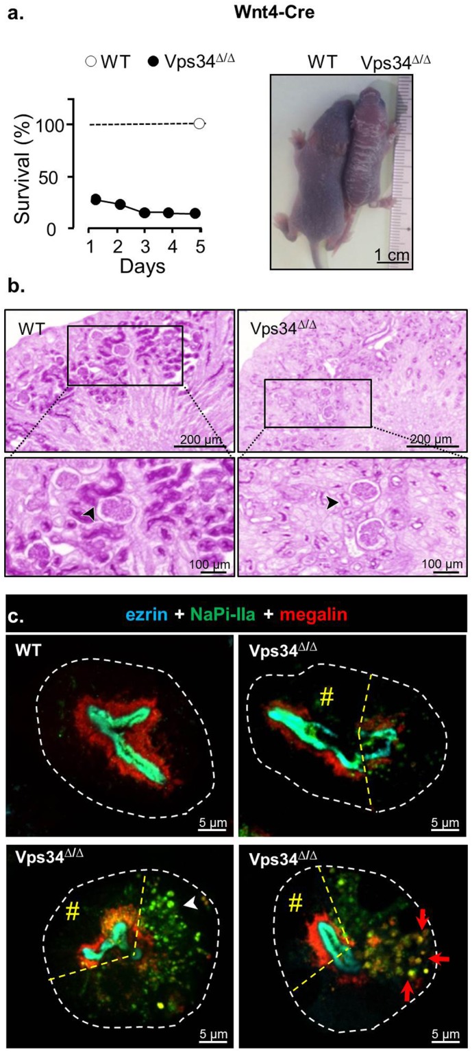
Figure 1.

Mouse model A. Wnt4-driven excision of Vps34 exon 21 (Wnt4-Vps34Δ/Δ) causes perinatal kidney lesions and death. (a) Survival. Offspring of Vps34fl/fl x Wnt4-Cre; Vps34fl/+ was genotyped within 24 h after birth and surviving mice were sacrificed at P5. At the left, the survival of Vps34Δ/Δ mice is expressed as percentage of WT littermates (n = 25 WT pups versus 5 Vps34Δ/Δ at P1 and 3 Vps34Δ/Δ at P5, euthanized at this age). At the right, a representative surviving Vps34Δ/Δ pup is compared with a WT littermate at P5. (b) Histology after PAS staining at P5. Boxed areas are enlarged below. Arrowheads point to apical tubular PAS staining. Note that Wnt4-driven excision alters kidney growth but is compatible with normal morphogenesis of glomeruli and kidney tubules, yet with much reduced apical PAS staining. (c) Confocal immunofluorescence at P5. WT (upper left) vs Vps34Δ/Δ kidney deep (mature) cortices were immunolabeled for brush border (ezrin, cyan), NaPi-IIa (green) and megalin (red). Contours of proximal tubules recorded by bright-field microscopy are delineated by dashed white lines. Three representative views from discrete (upper right), moderate (lower left) to extensive (lower right) mis-localization of (sub)apical membrane proteins in deep cytoplasm of some PTCs contrasting with preserved localization in adjacent cells of the same tubular section shown at left of dashed dashed lines (#, mosaicism). Notice stratification in mature WT PTCs between pale green layer (full co-localization of ezrin and NaPi-IIa at brush border), and subapical red layer showing major megalin pool, with “hairy” deeper extensions. In altered Vps34Δ/Δ PTCs, preferential mislocalization of NaPi-IIa yields green dots (lower left, arrowhead); shared mis-localization of NaPi-IIa and megalin generates yellow dots (lower right, red arrows). Whole cortical views are shown in Supplementary Fig. 2.
