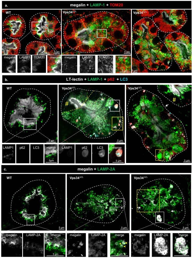
Figure 7.
Abrogation of macro-autophagy in Pax8-Vps34Δ/Δ PTCs. (a) Mitophagy. In WT PTCs, megalin (white), late endosomes/lysosomes (LAMP-1, green) and mitochondria (TOM20, red) define distinct layers. In Vps34Δ/Δ PTCs, note the loss of stratification (central panel, right to dashed yellow line; right panel, below dashed yellow line). TOM20 is never detected in mislocalized late endosomes/lysosomes. (b) Selective p62-dependent autophagy. In WT PTCs, LAMP-1-labeled late endosomes/lysosomes (green) in subapical zone are segregated from the LT-lectin-labeled apical cytoplasmic layer (white). No p62 (red) and LC3 (blue) puncta is visible. Vps34Δ/Δ PTCs sections usually show several punctae double-labeled for p62 and LC3 in the basal cytoplasm (compare with preserved cell indicated by yellow #). For P7, see Supplementary Fig. 7. (c) Chaperone-mediated autophagy. In WT PTCs, LAMP-2A labels similar structures as LAMP-1, polarized in the subapical cytoplasm, and likewise segregated from megalin. In Vps34Δ/Δ, LAMP-2A-surrounded structures are much more abundant, extend into basal cytoplasm where they are often enlarged (arrows in right panel; area boxed in yellow) and become labeled for megalin (arrowheads in central panel). The enlarged boxed area shows lysosomal lumen filled by LAMP-2A-labeled objects.

