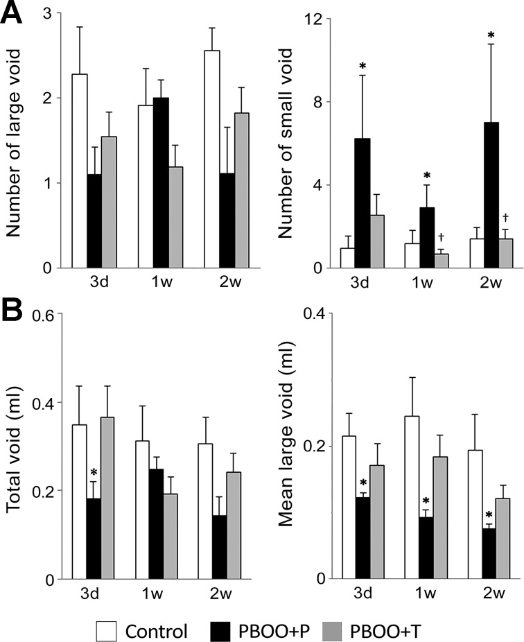
Fig. 7.
Micturition pattern analyses in spontaneous void spot assays. Micturition patterns were assessed on 3 days and 1 and 2 wk after the surgery. A: number of the urine spots of large void (≥50 μl, left) and small void (<50 μl, right). B: volume of total void (left) and mean volume of the large void spots (right). Values are means ± SE; n = 10–12 per group. *P < 0.05, vs. control group; †P < 0.05, between PBOO + P and PBOO + T groups.

