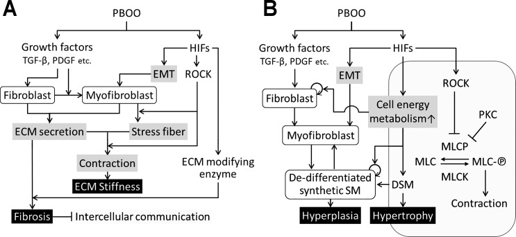
Fig. 9.
Schematic model of hypothetical mechanisms involve in PBOO-induced pathology in bladders. PBOO activates 2 major signaling pathways; growth factors including TGF-β, PDGF, and HIFs. Growth factors contribute to PBOO-induced pathogenesis through activation of fibroblast and myofibroblast, while HIFs activate genes in different types of cells, involving in multiple stage of bladder pathology secondary to PBOO. A: ECM remodeling through 2 signaling pathways leads to fibrosis and bladder stiffness. B: an enhancement of cell energy metabolism in the activated fibroblast, myofibroblast, synthetic smooth muscle, and DSM result in DSM hyperplasia/hypertrophy. Inhibition of MLCP by ROCK and PKC promotes sustained contraction of DSM. ECM, extracellular matrix; EMT, epithelial mesenchymal transition; SM, smooth muscle; DSM, detrusor smooth muscle; MLC, myosin light chain; MLC-℗, phosphorylated myosin light chain; MLCP, myosin light chain phosphatase; MLCK, myosin light chain kinase; TGF-β, transforming growth factor-β.

