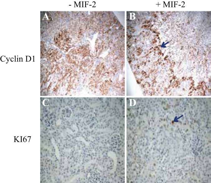
Fig. 6.

Immunohistochemistry (IHC) staining for cell proliferation markers in mouse kidney tissue 48 h after severe I/R injury. A: IHC for cyclin D1 in cortical tissue of Mif−/− mice without treatment. B: IHC stain for cyclin D1 in Mif−/− mice treated with MIF-2/D-DT. There is robust cyclin D 1 staining in proximal tubule at the cortico-medullary junction (blue arrow). C: IHC stain for Ki-67 in cortical tissue of Mif−/− without treatment. D: IHC staining for Ki-67 in Mif−/− mice, treated with MIF-2/D-DT, shows strong Ki-67 staining in proximal tubule cells (blue arrow); n = 6–8 animals per group.
