Fig. 3.
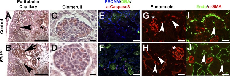
Flk1ST−/− mutant kidneys have dilated peritubular capillaries. A and B: immunohistochemistry staining for Endomucin (brown) at E15.5 show controls (A) with normal small caliber microvessels; however, mutants (B) displayed dilated peritubular capillaries (arrowheads) and a lack of organization (arrows) although the glomerular capillaries (stars) were unaffected. C and D: hematoxylin and eosin (H&E)-stained glomeruli at E18.5 reveal no obvious differences between control (C) and mutant (D) glomerular capillaries. E and F: control (E) and Flk1ST−/− kidney sections (F) stained for PECAM (endothelial cells, blue), DBA (collecting ducts, green), and activated caspase 3 (apoptosis, red) reveal markedly more apoptotic nuclei in mutant endothelial cells that surround the collecting ducts than in controls (E). G and H: postnatal day 30 (P30) kidneys stained with Endomucin (microvasculature, red) that shows significant dilatation in the mutant (H) peritubular capillaries (arrowheads) compared with controls (G), while glomerular capillaries appear equivalent (asterisk). I and J: 30 kidneys stained with Endomucin (green) and costained with the smooth muscle marker α-smooth muscle actin (α-SMA; red). The large muscular vessels show abundant α-SMA (red) in both the controls (I) and mutants (J). However, the mutant peritubular capillaries are dilated and do not express α-SMA (arrows). Scale bars: A and B = 200 μm; C and D = 25 μm; E–J = 100 μm.

