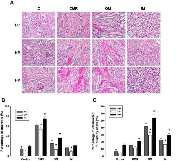
Fig. 6.
Histology. A: histology by PAS staining showed slices of cortex, CMR, OM, and IM. Kidney injury was quantitatively measured by percentage of tubular necrosis (B) and obstructed by tubules casts (C) in the cortex, CMR, OM, and IM in AKI groups by clamping renal veins with low renal artery pressure (LP), normal renal artery pressure (NP), and high renal artery pressure (HP) (n = 6). *P < 0.05 vs. LP group and HP grout. Quantitative analysis was performed on 10 randomly chosen fields in each kidney tissue slice.

