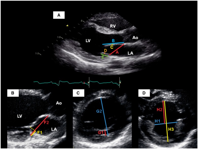
Figure 1.
Echocardiographic measurements in the parasternal long-axis view (A and B), and parasternal short-axis views of the left ventricle at the level of the mitral valve (C) and papillary muscles (D). A, mitral annular diameter; Ao, aortic root; B, anterior leaflet length; C, anterior leaflet thickness; D, posterior leaflet thickness; E, posterior leaflet length; F1, distance from posterior mitral hinge point to leaflet coaptation; F2, mitral annular diameter at end-systole; G1, distance from posterior wall of LV to posterior MV leaflet; G2, LV internal diameter at this level; H1, medio-lateral papillary muscle separation; H2, papillary-septal separation; H3, LV internal diameter at this level; LA, left atrium; LV, left ventricle; RV, right ventricle.

