Figure 2.
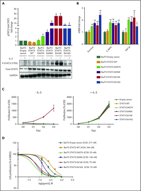
Novel STAT3 mutations cause constitutive STAT3 activity and are sensitive to pharmacologic inhibition. (A) Western blot analysis of pSTAT3 (Y705) and total STAT3 protein expression level in Ba/F3 cells expressing empty vector, STAT3WT, and novel STAT3 mutants after culture for 6 hours in medium with and without IL-3. Bands were quantified with Image J, and protein expression levels were represented as fold change of pSTAT3/total STAT3 relative to empty vector. *P < .05 compared with STAT3WT in medium without IL-3; #P < .05 compared with STAT3WT in medium with IL-3. (B) mRNA expression of STAT3 target genes in Ba/F3 cells expressing empty vector, STAT3WT, and novel STAT3 mutants after culture for 6 hours in medium without IL-3. Results were represented as fold change relative to empty vector and normalized against housekeeping gene NONO. *P < .05 compared with empty vector; #P < .05 compared with STAT3WT. (C) Cell viability assays of Ba/F3 cells expressing empty vector, STAT3WT, and novel STAT3 mutants with and without IL-3 up to 72 hours. (D) Cell viability assays with dimethyl sulfoxide vehicle and Stattic (0.125 µM, 0.25 µM, 0.5 µM, and 1.0 µM) for 72 hours in empty vector, STAT3WT, and novel STAT3 mutant Ba/F3 cells. All results are expressed as mean ± SD of 3 independent experiments.

