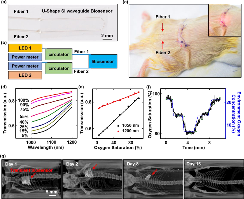
Figure 6. Monitoring of blood oxygen saturation in a live animal model using a transient Si waveguide-based biosensor.
(a) Image of a transient biosensor. Two bioresorbable fibers connect to a U-shaped Si waveguide bonded to a film of PLGA with the its top surface exposed. (b) Schematic illustration of the photonic operation principle of the transient biosensor: Each of the two bioresorbable fibers (labeled Fiber 1 and Fiber 2), probing externally from the transient biosensor, connects via a fiber circulator to a light source (1050 nm wavelength for Fiber 1, and 1200 nm wavelength for Fiber 2) and a power meter for delivering and detecting light, respectively. (c) Image of a transient biosensor implanted into the subcutaneous region near the thoracic spine of a mouse. Inset: image of red light (wavelength: 660 nm) passing into the biosensor. (d) In vitro measurements of oxygenation of hemoglobin in phosphate-buffered saline (PBS) by near infrared absorption measurements using a transient biosensor. Hemoglobin concentration: 1.5 g/dL. The amount of sodium dithionite in the solution defines the oxygenation level, across a relevant range. (e) Calibration curves corresponding to absorption at 1050 nm and 1200 nm measured in vitro. (f) In vivo measurements of blood oxygen saturation as a function of time during changes in the concentration of oxygen (labeled in blue) in the surrounding environment. Results measured by the transient biosensor and commercial oximeter are labeled in black and green, respectively. (g) Computed X-ray tomography (CT) image (processed by volume rendering analysis) of the transient biosensor implanted into a mouse at various stages of implantation from day 1 to day 15. The red arrow highlights the location of transient biosensor, which is no longer visible under CT on Day 15. All the CT images share the same scale bar.

