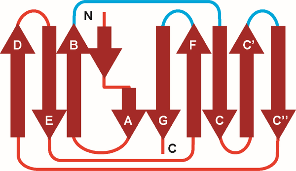
Figure 1.
2D topology diagram of immunoglobulin light chain (LC) fold. The nine β-strands that form theframework regions. These strands are connected by unstructured loops in a Greek key pattern. The loops (blue lines) that connect strands B and C, C’, C”, and F and G are determine the specificity of the antigen-antibody interactions, and are known as the complementarity-determining regions (CDRs).

