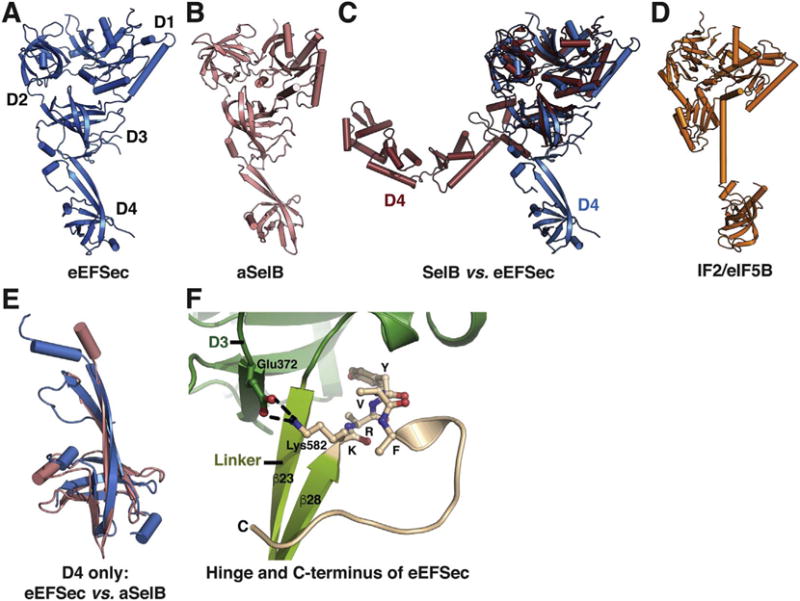
Fig. 3.

Conservation and divergence of Sec elongation factor structures. The structure of eEFSec (A) resembles that of aSelB (B) in spite of slight divergence of D4. Conversely, analogous comparison with SelB (C) reveals that the structural conservation is preserved within D1–3, whereas D4 adopts both the distinct structures and orientations. A cartoon representation of IF2/eIF5B (D) confirms its phylogenetic relationship with eEFSec and SelB. (E) Structural overlay of D4 from eEFSec (blue) and aSelB (pink) reveals slight differences between the domains. (F) The conformation of the extreme C-terminus (beige) of eEFSec and its interactions with linker (light green) and D3 (green). (For interpretation of the references to colour in this figure legend, the reader is referred to the online version of this chapter.)
