
Figure 3. Skull release more of cells after stroke.
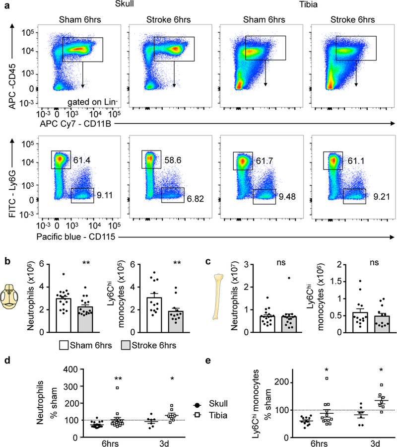
a, Representative flow cytometry plots of skull and tibia bone marrow 6 hours after stroke induced by 30 min tMCAO or sham controls (6 experiments). Additional gating is shown in Supplementary Fig. 2. b, Neutrophil and monocyte numbers in skull after stroke or sham controls. Data are mean ± s.e.m.. Neutrophils, n=16 stroke, n=17 sham, 6 experiments; Ly6Chi monocytes, n=13 stroke, n=12 sham, 4 experiments; two-tailed Mann-Whitney test, neutrophils, **P=0.008; monocytes, **P=0.007. c, Neutrophil and monocyte numbers in both tibiae. Data are mean ± s.e.m.. Neutrophils, n=16 stroke, n=17 sham, 6 experiments; Ly6Chi monocytes, n=13 stroke, n=12 sham, 4 experiments; two-tailed Mann-Whitney test, neutrophils, P=0.49; monocytes, P=0.54. d,e, Data normalized to sham at 6hrs (neutrophils, n=16 and monocytes, n=12 per condition; 4 experiments) and 3 days after stroke (n=7 per condition, 3 experiments) and displayed as mean (center) ± s.e.m. (error bars). Two-tailed paired Wilcoxon test, neutrophils, 6hrs, **P=0.002 skull versus tibia; 3 days, *P=0.031 skull versus tibia; monocytes, 6hrs *P=0.016 skull versus tibia; 3 days,*P=0.016 skull versus tibia. See Supplementary Fig. 5 for spine.