
Figure 7. Electron microscopy of channel.
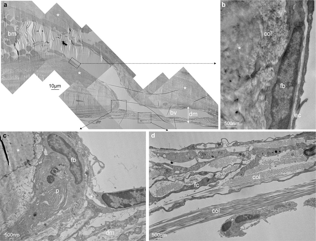
a, Entire channel connecting a skull bone marrow cavity (bm) filled with blood cells with a blood vessel (bv) in the dura mater (dm) while traversing the inner bone cortex (asterisks), single experiment. b, Channel is clad with endothelial cells (ec), above fibroblasts (fb). c, Connective tissue in the dura mater (dm) in the vicinity of the channel, fibroblast (fb), plasma cell (p). d, Dura mater with collagen (col) and fibrocyte (fc).