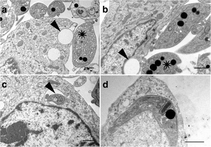
Fig. 10.
Transmission electron micrographs of potoroo kidney epithelial cells (PtK2) after incubation with Trypanosoma copemani G2 for 48 h. a Parasite attached to the surface of a cell. Parasite indicated by an asterisk is attached to the surface of the cell with a vacuole (arrow) underneath. b Two parasites attached to the surface of a cell one parasite (asterisk) with a vacuole underneath (arrow). c T. copemani inside a cell, inside a vacuole recognisable by the flagellum (arrow). d T. copemani attached to a cell, recognisable by the kinetoplast (arrow). Scale-bars: 1 μm

