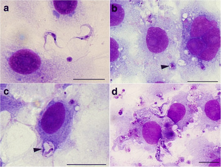
Fig. 3.
Microscopy of Trypanosoma copemani G2 incubated with potoroo kidney epithelial (PtK2) cells. a Light microscopy of G2 trypomastigotes. b Light microscopy of G2 amastigote-like cells inside PtK2 cells (arrowhead). c G2 trypomastigote inside a vacuole within the cell (arrowhead). d G2 attached to the outside of cells. All images are stained with Diff-Quik. Scale-bars: 20 μm

