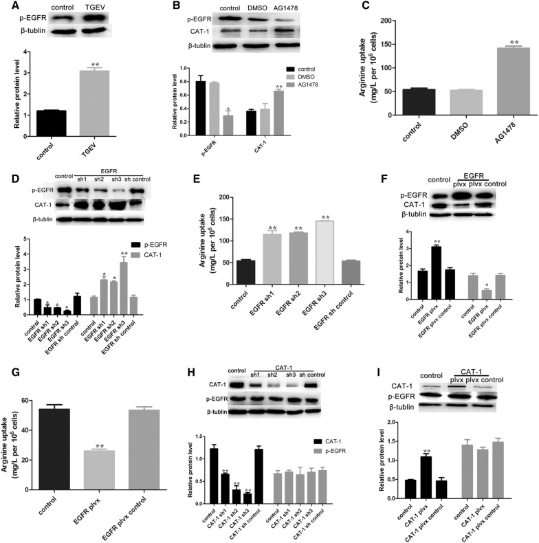
Figure 4.
TGEV infection decreases arginine uptake via activated p-EGFR. A Western blot analysis was performed to determine levels of p-EGFR at 48 hpi. B TGEV-infected cells were treated with PBS, DMSO (100 nM), or AG1478 (100 nM) for 12 h, and then were analyzed to determine protein expression levels of p-EGFR and CAT-1. C Arginine concentrations in the culture medium from each group were assayed. D Cells were transfected with EGFR specific shRNAs or the shRNA control for 48 h. p-EGFR and CAT-1 expression were evaluated by Western blot analysis. E Culture medium from each group was assayed to determine arginine uptake. F Cells were transfected with a plvx-EGFR shRNA or the plvx control, and analyzed for p-EGFR and CAT-1 protein levels by Western blot. G The uptake of arginine was assayed. H Cells were transfected with three CAT-1 specific shRNAs or the shRNA control, and analyzed for CAT-1 and p-EGFR protein levels by Western blot. I Cells were transfected with a plvx-CAT-1 shRNA or the plvx control for 48 h, and analyzed for CAT-1 and p-EGFR protein expression by Western blot. Differences were considered significant at *0.01 < p < 0.05, **p < 0.01.

