
Figure 1.
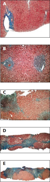
Progression of liver fibrosis as shown on a series of biopsy specimens and as classified by the Metavir schema. A: Liver with no fibrosis. B: Stage 1, fibrous expansion into some portal areas. C: Stage 2, fibrous expansion in most portal areas, with occasional portal-to-portal bridging. D: Stage 3, fibrous expansion of portal areas with marked bridging, including portal-to-portal and portal-to-central bridging. E: Stage 4, cirrhosis. Adapted with permission from Gregory T. Everson, MD, University of Colorado Denver.