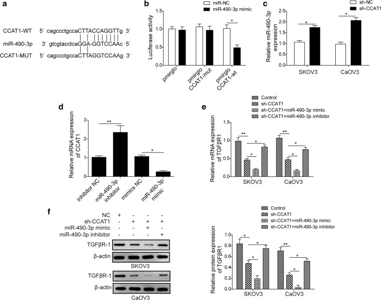
Fig. 3.
LncRNA CCAT1 directly targets miR-490-3p. a Diagram of bioinformatic prediction of binding site of miR-490-3p by CCAT1. b Cells were co-transfected with miR-490-3p mimics and wildtype CCAT1 or mutant CCAT1luciferase reporter plasmid. The cell lysates were harvested for luciferase assay. c RT-qPCR analysis showed the level of miR-490-3p in sh-NC (scramble) and sh-CCAT1 (CCAT1 knockdown) ovarian cancer cells (SKOV3 and CaOV3). d The level of CCAT1 of ovarian cancer cells which transfected with miR-490-3p mimic and miR-490-3p inhibitor was detected by RT-qPCR. e RT-qPCR analysis showed the mRNA level of TGFβR1 in ovarian cancer cells (SKOV3 and CaOV3) transfected with sh-NC (scramble), sh-CCAT1 (CCAT1 knockdown), sh-CCAT1 (CCAT1 knockdown) plus miR-490-3p mimics, sh-CCAT1 (CCAT1 knockdown) plus miR-490-3p inhibitor. f The protein level of TGFβR1 in ovarian cancer cells (SKOV3 and CaOV3) transfected with sh-NC (scramble), sh-CCAT1 (CCAT1 knockdown), sh-CCAT1 (CCAT1 knockdown) plus miR-490-3p mimics, sh-CCAT1 (CCAT1 knockdown) plus miR-490-3p inhibitor was measured by western lot assay. All data were represented as mean ± SD from three biological replicates (*P < 0.05; **P < 0.01)

