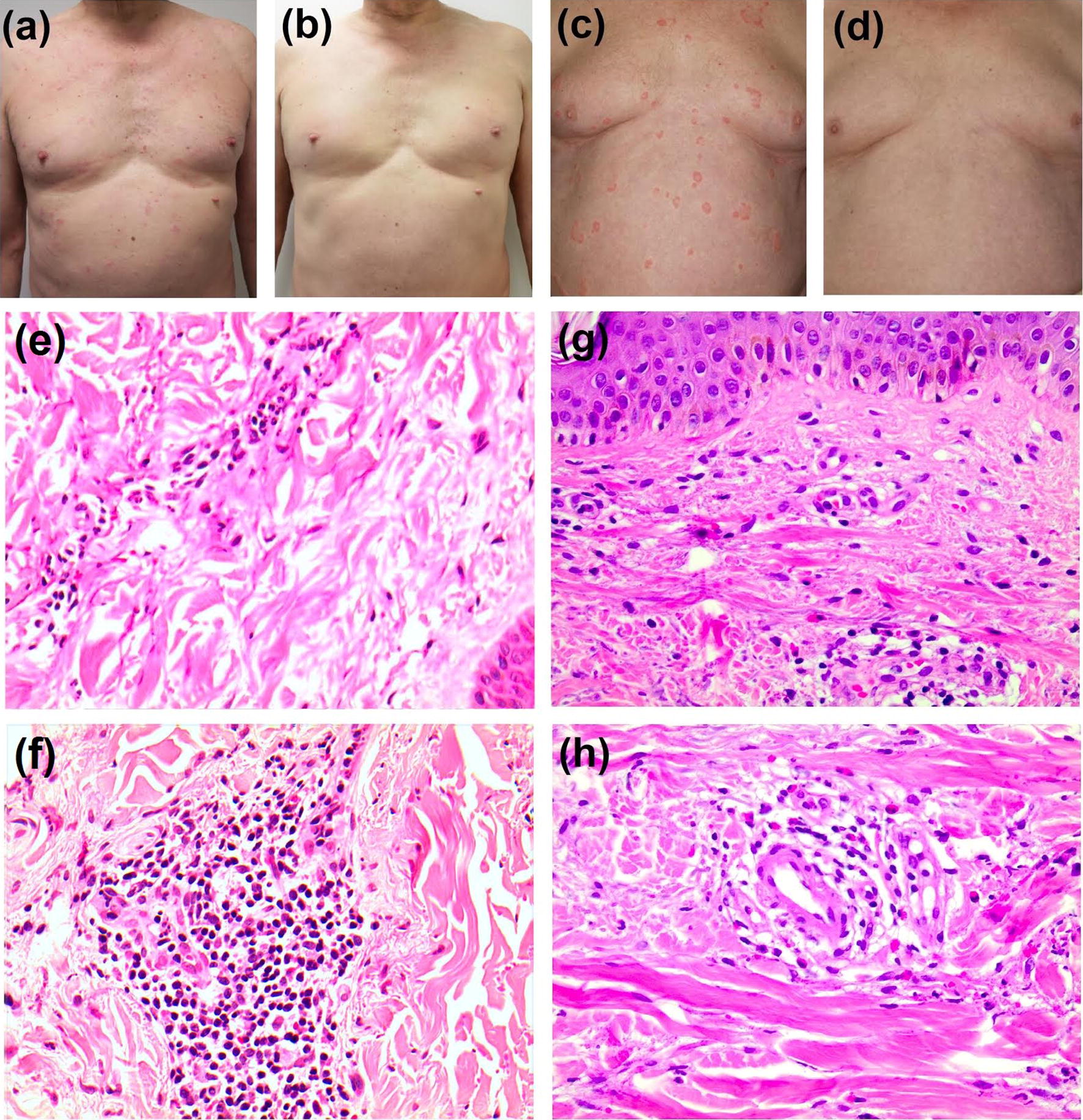
Fig. 1.

Patient 1—clinical photography of patient before (a) and after (b) treatment, histopathological sections of skin showing leukocytoclastic vasculitis with mild erythrocyte extravasation (e, f). Patient 2—clinical photography of patient before (c) and after (d) treatment, and histopathological sections of skin showing leukocytoclastic vasculitis with perivascular neutrophilic infiltrate and erythrocyte extravasation around small dermal vessels (g) and fibrinoid deposits within a vessel wall (h)
