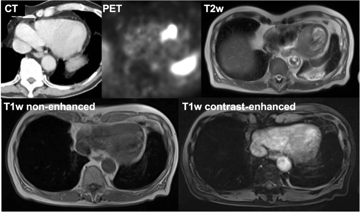
Fig. 3.
Appearance of a myocardial metastasis on liver MRI performed as part of the complementary staging of the NET disease. A 72-year-old male patient with a G1 neuroendocrine tumor of the small intestine demonstrates a myocardial metastasis in the interventricular septum (evident on CT imaging) with strong 68Ga-DOTATATE tracer uptake in the PET image. The morphologic appearance in the complementary liver MRI is characterized by an intermediate T2w signal and isointense signal on non-enhanced and contrast-enhanced T1w images

