Fig. 2.
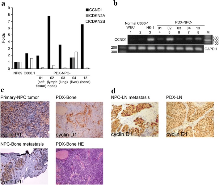
CCND1 mRNA expression and IHC staining in NPC patients and PDX tumors. (a) The expression fold change of candidate genes (CCND1, CDKN2A and CDKN2B) are indicated based on the cDNA microarray data of five PDX tissues, and C666–1 (EBV-positive NPC cells) and NP69 (immortalized normal nasopharyngeal cells, as control) cell lines. (b) Agarose gel electrophoresis of RT-PCR products of CCND1 in PBMC, two NPC cell lines and five PDXs (GAPDH serves as an internal control). Cyclin D1 IHC staining in (c) NPC no.13 patient, with NPC primary site, NPC metastatic to bone, and PDX-Bone tumor and (d) NPC no.2 patient, with NPC metastatic to lymph node, and PDX-LN tumor

