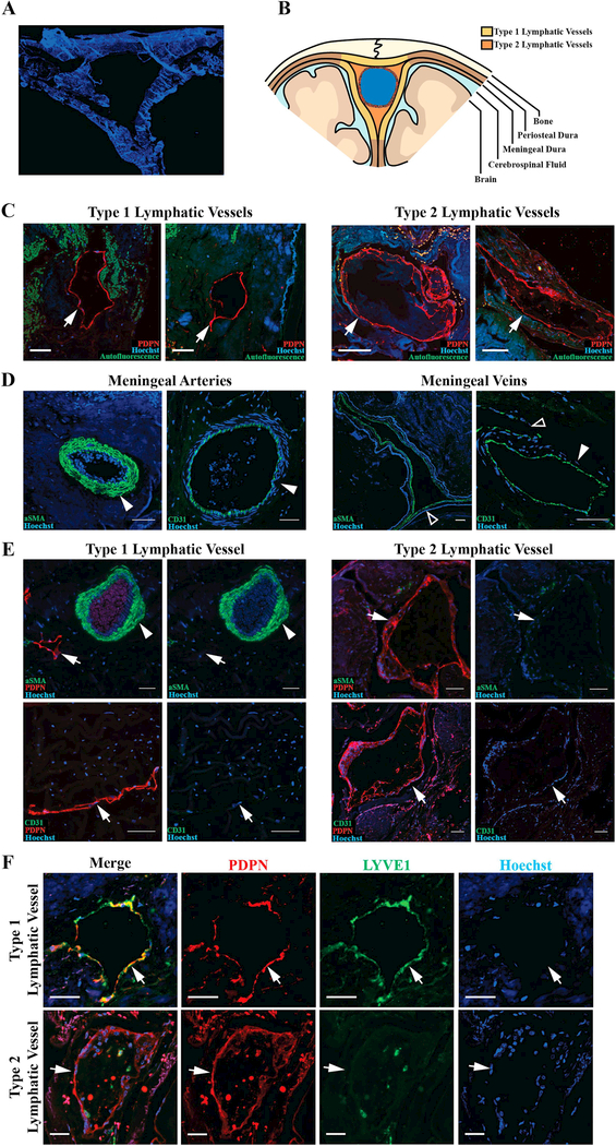
Figure 1. Lymphatic vessels with variable morphology invest the human meninges.
A. Representative image of coronal section of human superior sagittal sinus and meninges. B. Schematic demonstrating regions of the meninges where lymphatic vessels were found. Yellow regions represent the general distribution of Type 1 vessels and orange regions represent Type 2 vessels. C. Representative images of Type 1 and 2 lymphatic vessels (arrows). D. Dural blood vessels, including arteries (solid arrowhead) and veins (hollow arrowhead), labeled with vascular smooth muscle cell marker alpha-smooth muscle actin (aSMA) and the blood endothelial cell marker CD31. E. Dural lymphatic vessels do not co-label with aSMA or CD31. F. Type 1 vessels are PDPN+LYVE1+ and Type 2 vessels are PDPN+LYVE1-. Scale bars in C and D represent 100 μm and bars in E and F represent 50 μm.

