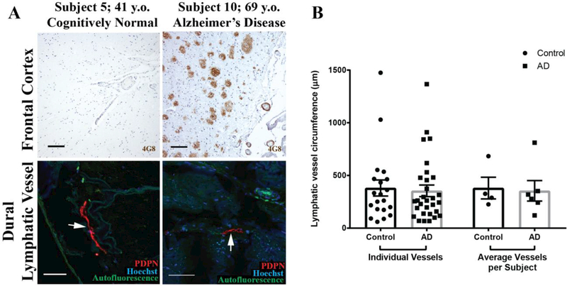
Figure 2. Meningeal lymphatic vessels in AD and control subjects.
A. Frontal cortical Aβ plaques and leptomeningeal vascular Aβ deposition in control and AD subjects. Type 1 and Type 2 meningeal lymphatic vessels (arrows) are readily detected among both groups. B. Quantification of lymphatic vessel circumference (14 Type 1 and 6 Type 2 vessels in control subjects; 22 Type 1 and 8 Type 2 in AD subjects). Columns on left reflect all vessels from all subjects (n = 20 and 30 from control and AD subjects, respectively). Columns on right reflect subject-wise averages of lymphatic vessel circumferences (n = 4 and 6 control and AD subjects, respectively). No group-wise differences in lymphatic vessel circumference were observed (unpaired two-tailed T-test with Welch’s correction, p= 0.78 and 0.85 for individual vessels and average vessels per subject, respectively).

