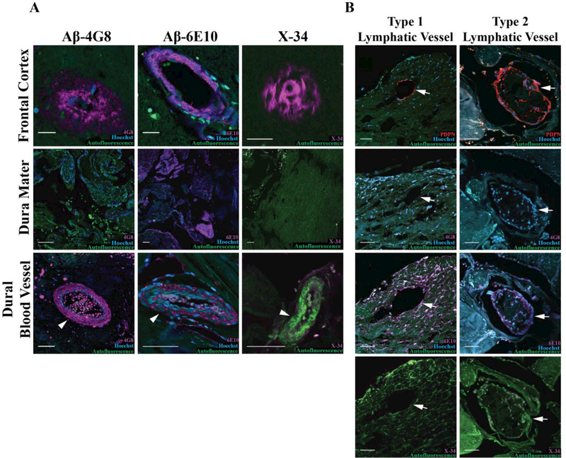
Figure 3. Dural lymphatic vasculature and meningeal Aβ immunoreactivity in AD subjects.
A. Detection of Aβ immunoreactivity with 6E10 and 4G8 clones, and Aβ aggregates with the congophilic X-34 dye in frontal cortex (top), within dural tissue (middle), and in meningeal blood vessels (bottom, arrowhead). B. Representative Type 1 (left) and Type 2 (right) lymphatic vessel (arrows). Sequential slices stained with PDPN, 6E10, 4G8 and X-34 shows that co-localization between PDPN and the 6E10 in some vessels. PDPN co-localization with 4G8 immunoreactivity or with X-34 labeling was not observed. Scale bars in frontal cortex micrographs represent 20 um and scale bars in other micrographs represent 50 μm.

