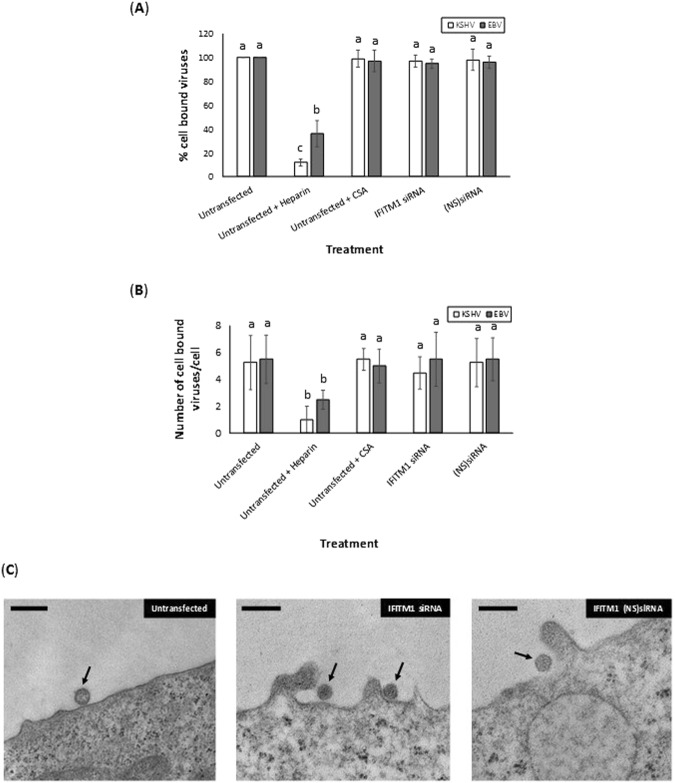
Figure 3.
IFITM1 enhancement of EBV and KSHV infection of cells is at a post-attachment stage of virus entry. (A) EBV and KSHV binding to BJAB cells were monitored in cells that were untransfected, untransfected and treated with heparin or CSA, or transiently transfected with IFITM1-specific siRNA, or transfected with non-specific (NS) siRNA. Data was plotted to represent the percentage of EBV or KSHV binding to BJAB cells treated differently compared to the untransfected cells. (B) EBV and KSHV were allowed to bind at 4 °C to BJAB cells that were untransfected, or transiently transfected with IFITM1-specific siRNA, or (NS) siRNA. Viruses incubated with soluble heparin and chondroitin sulfate served as controls. After 60 min, cells were fixed with 2% glutaraldehyde. Thin sections were examined by transmission electron microscopy. Enveloped virus particles bound to the cell surfaces of randomly selected 10 cells were counted by three different individuals using three different grids of their choice and the numbers were averaged. (C) Representative electron micrographs of KSHV binding to BJAB cells is depicted. Enveloped virus particles bound to the BJAB cells surfaces are indicated by the arrows. Magnification: 82,000 × (solid bar denotes 500 nm). Bars (A) represent average ± s.d. of five individual experiments. Columns (A,B) with different alphabets indicate the values to be statistically significant (p < 0.05) by LSD.

