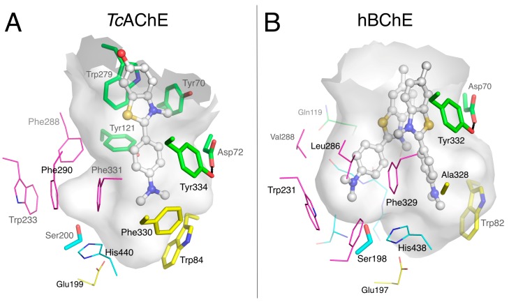
Figure 4.
Complexes of thioflavin T with TcAChE (A; pdb 2j3q) and hBChE (B; pdb 6esy). The gorge is depicted by its molecular surface (semi-transparent gray). Nitrogen atoms are in blue, oxygen atoms in red, sulfur atoms in yellow. The ligand is represented in ball and stick with carbon atoms in white. The residues in the vicinity of the ligand are represented in stick or lines. In the A-site, catalytic and oxyanion hole residues are in cyan, choline-binding pocket residues in yellow and acyl-binding pocket residues in magenta. P-site residues are in green.

