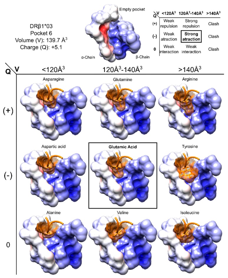
Figure 4.
Pocket volume and charge showing aa having different volumes and charge fitting into HLA-DRβ1*03 P6. Pocket characteristics and interactions are summarised at the top of the Figure, while the surface structures of aa occupying P6 are shown at the bottom. P6 surface is coloured according to its electrostatic potential set from −3 kT/e (negatively charged, red) to 3 kT/e (positively charged, blue). Figure based and adapted from [187]. Reproduced with permission from Patarroyo ME, Biochem. Biophys. Res. Commun.; published by Elsevier, 2017.

