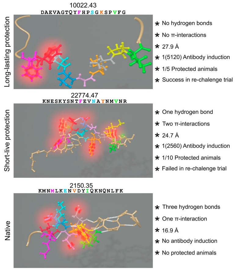
Figure 5.
Intramolecular forces' influence: the peptide’sequence and 3D-structure is shown on the left, represented by balls and sticks. Residues, fitting into HLA-DRβ PBR coloured the same as in Figure 3: H-bonds in white spheres, π-systems in blurred red and interacting hydrogens in blurred green. Structural and immunogenic features of the nine residues from cHABPs and mHABPs interacting with the PBR are shown on the right. Figure based and adapted from [253]. Reproduced with permission from Patarroyo ME, Biochem. Biophys. Res. Commun.; published by Elsevier, 2017.

