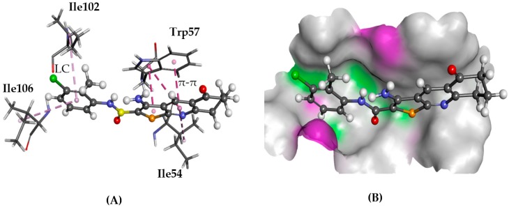
Figure 12.
The docked configuration of 1 to the binding site of NPSRb1 using GS scoring function. (A) π-π stacking and lipophilic contact (LC) are depicted as purple lines between the ligand and the amino acids; (B) 1 shown in the binding pocket with the protein surface rendered. Purple represents hydrogen bond donor regions, and green depicts hydrogen bond acceptor regions.

