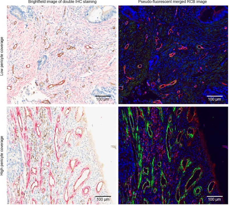
Figure 1.
Double IHC staining and image analyses of mCRC tissue.
Notes: Tissue sections were stained with anti-CD31 antibody (brown in the brightfield IHC images, red in the pseudo-fluorescent merged RGB images) and anti-α-SMA antibody (red in the brightfield IHC images, green in the pseudo-fluorescent merged RGB images). Brightfield and pseudo-fluorescent RGB images are shown to highlight the intensity of the staining. Scale bars, 100 µm.
Abbreviations: IHC, immunohistochemical; mCRC, metastatic colorectal cancer; CD31, cluster of differentiation 31; RGB, red-green-blue; α-SMA, alpha-smooth muscle actin.

