FIGURE 2.

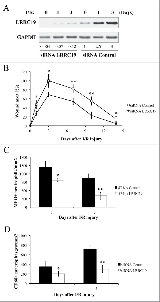
Knocking down LRRC19 reduces pressure ulcer formation and decreases the inflammatory cell infiltration in wound area. A) Western blot analysis of LRRC19 protein expression at the wound site during I/R injury. The wound area was topically applied with 100 pmol of either LRRC19 siRNA or control siRNA immediately after 1st ischemia delivery, followed by day 0, 1, 3, 6, 9, 12 after I/R injury induced. The end of 2nd ischemia was assigned day 0. The data are representative of n = 4 subjects for each group. The band intensity of LRCC19 was normalized against GAPDH. The normalized band densitometry of the siRNA control treated sample at day 0 was set as baseline of value 1. B) The average size of the wound area in mice (n = 6 for each group) treated with either LRRC19 or control siRNA during I/R injury were measured at day 0, 1, 3, 7 10 and 14 post injury. The end of 2nd ischemia was assigned day 0. The size of the ulcer in control group on day 3 after I/R was assigned a value of 100%. Immunohisochemistry was used to study the number of C) neutrophils (myeloperoxidase positive) and D) macrophages (CD68 positive) at the lesion site. Values were determined in six random microscopic fields (∼20,000 mm2) in n = 6 mice per group. The results are presented as cells per mm2. The data are presented as the mean ± SD (n = 4 for each time point). *p < 0.05, **p < 0.01, significantly different from control group.
