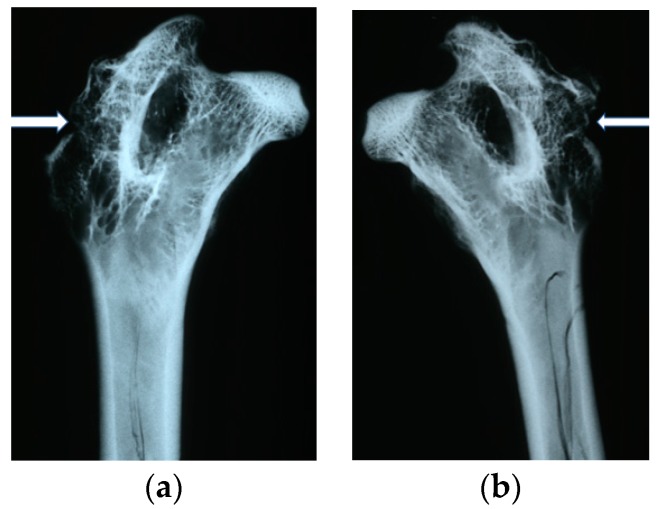
Figure 5.
Radiological images after implantation of the tested materials: (a) PLGA after six months. Traces of the implant channel are visible, filled with thin bone trabeculae. The implant channel is closed with a thin bone cap. (b) PLGA + IGF1 after six months. In contrast to the opposite side, the implant channel is invisible, filled with numerous thick bone trabeculae, and there is a thicker bone cap at the point of the borehole.

