Figure 3.

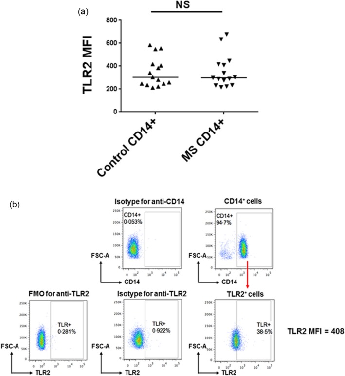
Toll‐like receptor (TLR)2 expression on CD14+ monocytes. (a) CD14+ monocytes from multiple sclerosis (MS) patients and controls were cultured for 4 h without stimulation and then stained with anti‐human CD14 and TLR2 antibodies. Mean fluorescence intensity (MFI) of TLR2 staining on CD14+ monocytes is shown. Medians are depicted. The results were analysed by Mann–Whitney test. (b) Representative flow cytometric plots for determining MFI of TLR2 staining on purified CD14+ monocytes.
